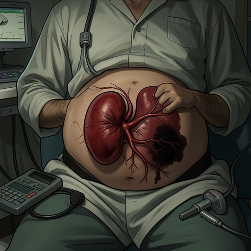
Proteinuria
Hepatitis C in Renal Disease, Hemodialysis and TransplantationProteinuria is the urinary manifestation that links hepatitis C virus infection to clinically significant renal injury in both chronic kidney disease and kidney transplantation. In HCV-positive renal allograft recipients, it is not merely a laboratory abnormality: it may be an early sign of chronic allograft damage, transplant glomerulopathy, or de novo immune complex-mediated glomerulonephritis, conditions associated with impaired graft survival and the need for close post-transplant surveillance.
Definition and Clinical Setting
Proteinuria denotes abnormal urinary protein loss and appears in the book as one of the principal renal consequences associated with chronic HCV infection. HCV is treated as a systemic disease rather than a disorder confined to the liver; it can induce renal disease, including immune complex-mediated glomerular lesions. Patients with HCV-associated glomerulonephritis are consistently HCV RNA-positive in serum, linking the renal finding to active viral infection rather than to antibody evidence alone. The distinction matters because anti-HCV antibodies indicate exposure, whereas detectable HCV RNA establishes active infection and helps define the context in which proteinuria should be interpreted.
The broader epidemiologic evidence also places proteinuria within a continuum of HCV-related kidney risk. In one study, chronic kidney disease was defined by either an estimated glomerular filtration rate below 60 ml/min/1.73 m2 or persistent proteinuria for more than six months. HCV-positive patients had a higher prevalence of chronic kidney disease than HCV-negative patients, 9.6% versus 5.1%, and developed both chronic kidney disease and end-stage renal disease more rapidly. A higher baseline viral load independently predicted chronic kidney disease. In another population analysis, HCV seropositivity was associated with albuminuria, particularly among people aged 40–59 years and those aged 60 years or older; among the older group, albuminuria occurred in 46% of HCV-positive individuals compared with 24% of those without HCV antibodies.
Proteinuria After Kidney Transplantation
After kidney transplantation, HCV-positive recipients have worse long-term patient and graft outcomes than HCV-negative recipients, and proteinuria is one of the pathways through which this disadvantage becomes clinically visible. HCV infection has been identified as an independent risk factor for post-transplant proteinuria. The mechanisms are not limited to one lesion: protein loss may arise from chronic allograft nephropathy, also described as chronic allograft nephropathy/interstitial fibrosis and tubular atrophy, from transplant glomerulopathy, or from HCV-associated glomerulonephritis developing in the renal allograft.
In biopsied HCV-positive recipients with proteinuria, chronic allograft nephropathy was the most frequent lesion, and nearly half of the anti-HCV-positive patients in one histopathological series had transplant glomerulopathy. The relationship between HCV and transplant glomerulopathy has not been entirely consistent across studies, but one comparison found anti-HCV antibodies in 33% of patients with transplant glomerulopathy versus 1.9% of those without it. A proposed explanation is that HCV may injure endothelial cells directly, indirectly, or through cytokines such as interferon, eventually producing the capillary and glomerular changes expressed clinically as proteinuria.
The association also intersects with immunology. HCV-infected recipients may have heightened cellular and humoral immune responses, and transplant glomerulopathy and glomerulitis are associated with anti-HLA antibodies and antibody-mediated rejection. Thus, proteinuria can reflect overlapping viral, autoimmune, and alloimmune injury rather than a single isolated cause. The book emphasizes that HCV remains an independent predictor of graft outcome even in protocol-biopsy studies, although the immunological risk profile of the HCV-infected population may contribute to the observed effect.
HCV-Associated Glomerular Disease
HCV-infected renal and liver allograft recipients are susceptible to immune complex-mediated glomerulonephritis, as are non-transplanted patients. De novo membranoproliferative glomerulonephritis was first reported in HCV-infected renal allograft recipients in 1995. Later observations identified type II mixed cryoglobulinemia, with an IgM-kappa monoclonal component, in patients who developed de novo HCV-related membranoproliferative glomerulonephritis after transplantation. Cryocrit levels in these allograft patients are usually very low, generally below 5%, which can make cryoglobulins difficult to detect and characterize; HCV RNA is often much more concentrated in the cryocrit than in serum, supporting a pathogenic role for the virus.
The clinical pattern of HCV-related membranoproliferative glomerulonephritis includes hypertension, microscopic hematuria, proteinuria, and renal dysfunction, sometimes accompanied by nephrotic syndrome. Biopsy may show mesangial proliferation, double contours, and parietal IgM and C3 deposits. Electron microscopy may be required to distinguish membranoproliferative glomerulonephritis from transplant glomerulopathy: the former has subendothelial electron-dense deposits, whereas the latter shows radiolucent areas in the glomerular basement membrane. The two lesions can coexist, demonstrating how immune-complex disease and alloimmune injury may operate together in the same graft.
Membranous glomerulonephritis is another possible explanation for proteinuria in an HCV-positive allograft. It is the most frequent type of de novo glomerulonephritis in HCV-negative renal allografts as well, and nephrotic syndrome is usually present. Unlike HCV-related membranoproliferative glomerulonephritis, however, membranous glomerulonephritis has no clearly established connection to HCV: cryoglobulins, immune complexes, and complement activation are usually absent. This contrast prevents proteinuria from being treated as proof of one particular HCV-mediated lesion.
Detection and Diagnostic Response
Because proteinuria may precede obvious allograft dysfunction, recommended follow-up after transplantation includes active surveillance for urinary protein loss alongside renal function, infection, deterioration of liver disease, new-onset diabetes after transplantation, cardiovascular disease, and neoplasia. The specific recommendation is to remain alert to proteinuria and/or microscopic hematuria. When either finding is present, a graft biopsy should be performed rather than attributing the abnormality automatically to HCV, chronic allograft injury, or rejection.
The biopsy is clinically important because the estimated prevalence of glomerular lesions in HCV renal allografts is approximately 5–15%, varying according to whether protocol biopsies or biopsies performed for cause are analyzed. Mild abnormalities and overlapping immunological damage can make pathological diagnosis difficult. Nevertheless, identifying de novo glomerulonephritis matters because it is associated with accelerated graft loss; recipients who have already lost a graft to de novo HCV-related glomerulonephritis may develop an earlier and more aggressive form in subsequent transplants.
When HCV-related glomerulonephritis is diagnosed, antiproteinuric medication should be started. The finding must also prompt consideration of active HCV infection and the competing severity of kidney and liver disease when antiviral treatment is evaluated. The AASLD guidance described in the book states that interferon-based antiviral therapy must be considered in people with chronic kidney disease, but treatment must be individualized according to kidney disease, liver disease, treatment risks, and comorbidities. Because the kidneys participate in the clearance of interferon and ribavirin, renal impairment alters treatment decisions; ribavirin is contraindicated when creatinine clearance is below 50 ml/min because accumulation can cause dramatic anemia.
Prognostic and Management Significance
Proteinuria is therefore both a manifestation and a warning signal. It can accompany HCV-related membranoproliferative glomerulonephritis, membranous glomerulonephritis, chronic allograft nephropathy, or transplant glomerulopathy, and several of these processes contribute to reduced death-censored graft survival. The increased graft-loss risk in HCV-positive recipients is also accompanied by higher mortality, driven by cardiovascular disease, liver disease, and serious infections. Proteinuria brings part of that otherwise diffuse risk into view at the level of the individual graft.
Despite these hazards, kidney transplantation remains the renal replacement therapy of choice for an HCV-infected patient with end-stage renal disease, because transplantation still provides benefit compared with other forms of kidney replacement therapy. That benefit depends on disciplined follow-up rather than on treating HCV status as a static label. Monitoring proteinuria, investigating it with graft biopsy when indicated, controlling urinary protein loss, managing immunosuppression carefully, and assessing active HCV and its extrahepatic complications together form the practical response to a finding that may mark the beginning of accelerated allograft injury.